 infobox@neuroproof.com
 +49 381 54345-660
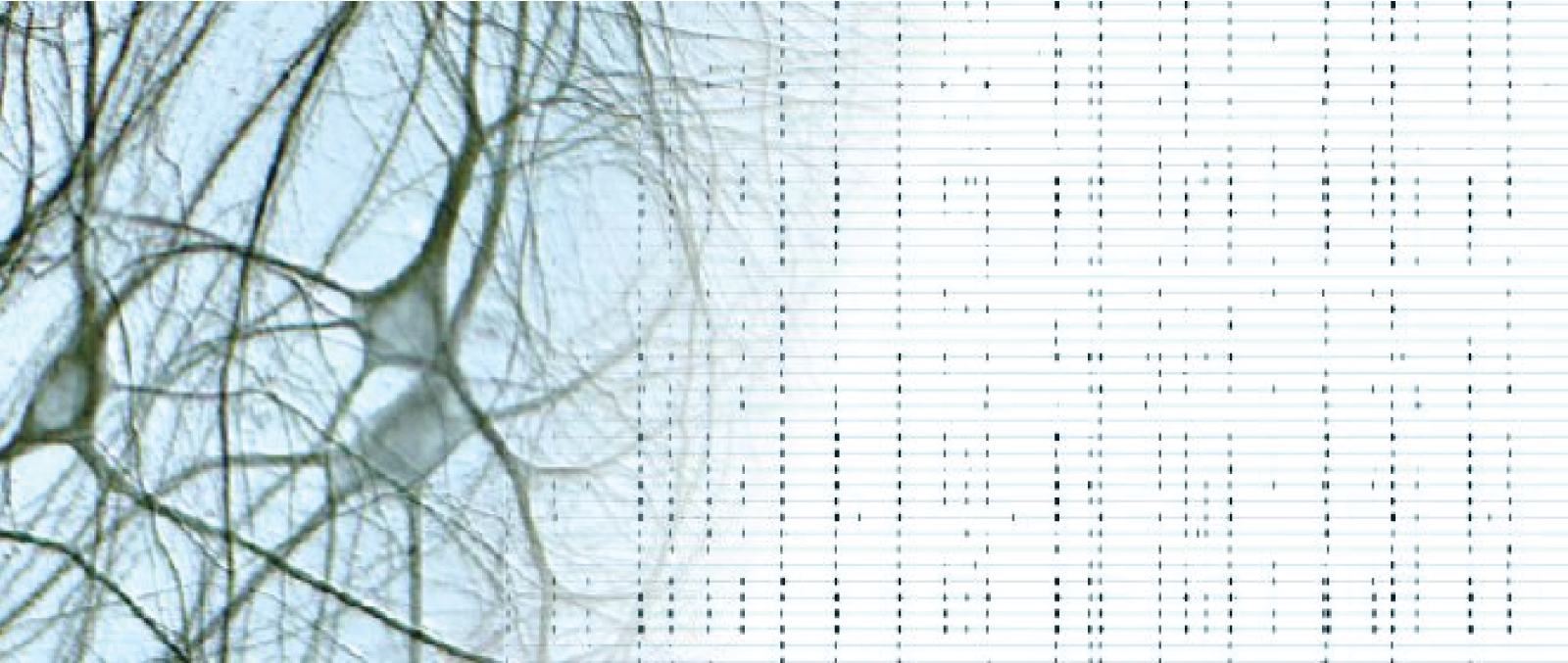
Primary Neuronal Cell Cultures
Primary neuronal cell cultures are best suited for drug discovery and drug development. They build complex systems as a co-culture of different cell types.
Advantages of primary neuronal cell cultures
Primary neuronal cell cultures freshly prepared from animals reproduce physiological conditions astonishingly similar to the in vivo situation. Therefore they are advantageous in their predictive capacity for drug screening and drug development. Physiological relevance compensates very often for the lack of non-human specificity. Primary neuronal cell cultures show a significant similarity in receptor composition to the in vivo situation. Prepared cultures from the early embryonic stages mimic neuronal development in high correlation to the in vivo situation.
Electrical functional primary neuronal cell cultures
Primary neuronal cell cultures on microelectrode arrays develop spontaneous electrical activity and generate extracellular action potentials. Extracellular action potentials present the natural function of neuronal cell cultures. Spontaneous electrical activity facilitates the physiological development of neuronal cell cultures, and it is a prerequisite for adequate physiological behavior, only with which a functional phenotypic screening is possible. Continuous action potential firing induces synapse formation and synaptic connectivity.
Comparison of primary neuronal cell cultures with human iPSC derived neurons
Human iPSC-derived neuronal cell cultures have clear advantages by ensuring that receptors are identical as in humans. More and more researchers test compounds in a human-based background. However, many open questions exist with human iPSC-derived neuronal cell cultures. From our experience, we see a more consistent physiological behavior in developmental processes in primary murine cultures than in human iPSC-derived neuronal cell cultures.
One another advantage of human iPSC-derived neuronal cell cultures taken from patients is the opportunity to create immediately in vitro disease models. NeuroProof has performed a range of drug screening projects with human IPSC derived neuronal cell cultures as patient-based disease models. Examples are aiming rare diseases like amyotrophic lateral sclerosis, ALS, spinal muscular atrophy, SMA, or fragile X syndrome.
Neurodevelopment in primary neuronal cell cultures
Primary neuronal cell cultures are prepared from progenitor cells at the embryonic stage. Mature and differentiated neuronal networks dissected at later stages can only be used as a complete tissue. The selection of the right embryonic day for preparation is essential for the optimal development of the cell culture. Progenitor cells are developing to a post-mitotic stage with a strong network of synaptic connections. These synaptic connections mirror the in vivo situation at a local base, so they present neuronal microcircuits with properties similar to the in vivo situation at the respective brain region of origin.
The evolution from progenitor cells to post-mitotic well-differentiated neuronal networks follows a precise developmental pattern. It is possible to determine the developmental day of a culture from its activity pattern with a precision of two days.
After four weeks in culture, the activity pattern stabilizes and is composed of one coordinated main burst pattern with several coordinated sub-patterns.
Furthermore, we have shown that specific receptors in comparison to others, change their expression in time as in the in vivo situation. For example, GABA-A receptors with the alpha1 subunit are expressed at low levels in the early phases and at higher levels after 28 days in vitro. For the GABA-A with alpha 2/3 subtype receptors, we notice a decreasing expression over time onwards.
Cell Types and receptor configuration
In primary neuronal cell cultures, neurons and glia cells, as astrocytes and microglia, are expressed similarly to the in vivo situation.
A mixture of excitatory and inhibitory neurons is responsible for excitatory and inhibitory activity balance, called E/I balance. Gabaergic neurons operate as inhibitory and glutamatergic neurons as the primary excitatory drivers, allowing studies of the E/I balance disturbance. The development from progenitor cells to post-mitotic well-differentiated neuronal networks follows a precise developmental pattern. It is possible to determine the developmental day of a culture from its activity pattern with a precision of two days.
After four weeks in culture, the activity pattern stabilizes and is composed of one coordinated main burst pattern with several coordinated sub-patterns.
Further, we have shown that specific receptors in comparison to others, change their expression as in the in vivo situation. For example GABA-A receptors with the alpha1 subunit are expressed at low levels in the early phases and higher levels after 28 days in vitro. The opposite for the GABA-A 2/3 receptors show a decreasing expression onwards over time.
Brain region-specific primary neuronal cell cultures
The right selection of the right brain region for drug discovery and drug profiling studies is essential. NeuroProof has developed preparation protocols for different brain regions and co-cultures of such regions.
Frontal cortex
The frontal cortex is dissected from the prefrontal cortex. Cells are prepared at embryonic day 16, E16. These cultures consist of approximately 20% neurons and 80% glial cells , including 1% microglia 28 days after plating. Frontal cortex cultures are used for the study of general toxicity, neurodevelopment, a wide range of diseases such as psychotic diseases and others.
Spinal cord with dorsal root ganglia
Neurons from the spinal cord are dissected at embryonic day 18 with and without dorsal root ganglia. These cultures contain capsaicin-sensitive neurons.
Spinal cord cultures with dorsal root ganglia are ideal for pain research and drug discovery.
Midbrain ventral
Midbrain cultures are dissected from the ventral midbrain at embryonic day 14, E14. The objective is the achievement of cultures containing dopaminergic cells, especially from the substantia nigra, the best possible configuration for studies targeting neurodegenerative diseases such as Parkinson’s disease.
Midbrain with nucleus basalis of Meynert
Midbrain cultures containing nucleus basalis of Meynert are used to study cholinergic neurons and cholinergic receptor activation. The nucleus basalis of Meynert is one of the most reach regions of cholinergic neurons. The culture is suited for all applications where the cholinergic pathway is of interest.
Hippocampus or hippocampal culture
The hippocampus is part of the limbic system. It is a heterogeneous but distinguishable structure. Preparation is usually at embryonic day 18. Preparations at earlier stages cause an epileptic-like behavior.
The hippocampus plays a vital role in the short term and long term. The hippocampus is essential for neurodegenerative diseases such as Alzheimer’s disease and others. NeuroProof is an expert in GABA-A alpha 5 subunit receptor studies with hippocampal cells.
Cerebellum
The cerebellum is a structured region of the brain. Preparation is at embryonic day 18, E18. To some extent, Primary neuronal cell cultures from the cerebellum contain oligodendrocytes.
Co-cultures
The combination of cells from different brain regions or the enrichment with specific cell types is often used to improve assay performance. For example, we can enrich cell cultures with microglia cells. Microglia cells have a dramatic influence on neurodevelopmental processes, which can be studied in assays with microglia enrichment.
Cell Culture Protocols
Dissection, dissociation, and plating of cells
Embryonic brain tissue is derived from gestating NMRI mice (Charles River, Sulzfeld, Germany). The tissue is dissociated enzymatically in DMEM or MEM with 10% horse and 10% fetal calf serum, including papain and DNase I. Cells are re-suspended in DMEM, containing 10μg/ml laminin. Cells are plated at a density of 7.5 × 10⁶ cells/ml, respectively, 150,000 cells per well.
Substrate preparation
Before plating MEAs, we use the 48 wells from Axion Biosystem, and they will be coated with freshly prepared 0.1% poly-ethyleneimine (PEI, Sigma, 181,978) dissolved in Borate buffer (Fisher Scientific, 28341). Alternatively, Poly-D-Lysin, PDL, can be used in combination with pretreatment of fibronectin or laminin.
Media
We use DMEM, or MEM for spinal cord cultures, containing 10% horse serum routinely. For serum-free cultivations, we use a neurobasal medium with a B28 supplement. Neuronal development is improved by the addition of the right supplements of amino and fatty acids and other components.
Maintenance
Culture conditions are 37◦C in a 10% CO2 atmosphere. Half-medium changes are performed twice per week. The developing co-cultures are treated on day 5 in vitro with 5-fluoro-2′-deoxyuridine to prevent glial proliferation and overgrowth.
Quality control
There is no better quality control than a functional screening of the primary neuronal cell cultures after their electrophysiological activity patterns are available. NeuroProof employs numerous quality parameters for the assessment of the quality of the primary neuronal cell cultures.